Engineering
Our project can be viewed as an overarching Design–Build–Test–Learn (DBTL) cycleA repeating engineering loop: design a solution, build it, test it, and learn from the result to improve the next design., progressing from the initial concept toward the envisioned integrated system of AnTigone. Within this broader process, several smaller DBTL cycles addressed specific experimental challenges, allowing us to iteratively refine individual components of our design.
Each cycle generated observations that informed subsequent experiments, prompted design modifications, and changed how we interpreted our results. As we had limited access and time in the lab, some DBTL cycles were carried out in parallel and some, mentioned in this section, are still open and we invite future researchers and students to carry out and build on what we assessed with our project.
Select a slice to read about it.
Our main challenges and small DBTL cycles refer to:
- Engineering Enzyme Specificity: making testosterone the most favorable substrate for our candidate enzymes.
- Construct Assembly: establishing a reliable cloning and verification strategy.
- Enzyme Production and Recovery: identifying suitable fusion tags and cultivation conditions for recombinant AKR1D1.
- Activity Characterization: developing reliable assays for evaluating testosterone-associated enzymatic activity.
In parallel, computational enzyme engineering informed our choice of variants for experimental investigation.
All cycles are interconnected: improvements in cloning allowed the production of alternative enzyme constructs, while expression and activity experiments revealed additional requirements for future optimizations.
Rational Engineering of Enzyme Specificity
Challenge
Our project aims to investigate testosterone transformation while limiting unwanted activity toward other substratesThe molecules an enzyme acts on and converts., particularly progesterone. Because these molecules share a similar steroid structure, modifying enzyme substrate preference represents an important engineering challenge.
Design
We investigated the active sites of human AKR1D1 (BBa_26ZLKFI1) and bacterial ci2350 (BBa_261I5W2C) to identify residuesThe individual amino acids that make up a protein. that could influence interactions with the C17 substituentsThe chemical groups attached to carbon 17 of the steroid, where testosterone and progesterone differ. of testosterone and progesterone.
Check out our designs and models.
Candidate point mutations were designed to favor testosterone binding or weaken progesterone interactions.
Build
We generated 24 computational AKR1D1 variants and 31 ci2350 variants. Structural stability was evaluated using PyRosettaA software package for modeling protein structures and predicting how mutations change their stability., and both testosterone and progesterone were independently dockedSimulated on a computer to predict how well a molecule fits into a protein's binding site. against variants that passed the selected stability threshold.
Thirteen AKR1D1 and seventeen ci2350 variants passed the initial stability screening.
Test
For AKR1D1, none of the stable variants showed a meaningful improvement in predicted testosterone selectivity beyond the uncertainty of the docking scoring function.
For ci2350, the R215A + V263I double mutant (BBa_262E3BYN) showed the strongest predicted shift toward testosterone, changing the selectivity scoreThe difference in predicted binding between testosterone and progesterone. More negative means a stronger preference for testosterone. from −0.1 kcal/molKilocalories per mole, a unit of energy. Here it measures predicted binding strength. in wild type to −0.9 kcal/mol, with a predicted stability score of +1.42 REURosetta Energy Units, the score software gives for how stable a protein structure is. Lower is more stable..
Check our results here.
Learn
The investigated AKR1D1 mutations did not produce meaningful predicted specificity improvements, suggesting that the selected residues were insufficient to discriminate between the two steroid substrates.
Improving the cloning workflow
Challenge
One of our main engineering challenges was assembling correct expression constructs for the candidate enzymes, AKR1D1 and ci2350. Our initial restriction-cloningCutting DNA with restriction enzymes at specific sequences and joining the pieces together to build a new construct. workflow using pET-11aA plasmid commonly used to produce proteins in E. coli. repeatedly produced low DNA recovery and unsuccessful transformations. In addition, incomplete restriction digestion could not be reliably identified because the NdeI and BamHITwo restriction enzymes, each cutting DNA at its own specific sequence. Together they excise a gene so it can be inserted into a plasmid. restriction sites are only approximately 40 base pairs (bp)The unit for measuring DNA length: one pair of matching DNA letters. apart.
Design
Our initial restriction-ligation strategy used the restriction enzymes NdeI and BamHI to cut the pET11-a plasmid in order to insert the genes. Following inconsistent results and unsuccessful attempts, we investigated whether plasmid purity, restriction efficiency, and vector-insert ligation preparation contributed to the cloning difficulties.
We subsequently redesigned the workflow using pET-BLANK, a pET-derived construct containing ci2350–MBP. Digestion was expected to release an approximately 3 kb insert, leaving a vector backbone of approximately 5.4 kb. Unlike the original strategy, this would allow the backbone to be separated from the released insert by agarose gel electrophoresis.
Build
Expand this section and find out about the iterative cycles!
Test
We improved first gel extraction yield and purity using different kits and we evaluated the resulting constructs through careful choice of controls and screening of samples to include in colony PCRA quick check of bacterial colonies for the right insert: a PCR run directly on a small sample of each colony., restriction digestion and sequencing. However, colony PCR still revealed unsuccessful cloning attempts. Further investigation suggested incomplete NdeI digestion. Because the two restriction sites in pET-11a were separated by only approximately 40 bp, conventional agarose gel electrophoresis could not reliably distinguish singly digested from fully digested vector.
In contrast, pET-BLANK digestion produced distinguishable backbone and insert fragments. Following the revised ligation procedure, colony-PCR-positive AKR1D1–SUMO and AKR1D1–TrxA candidates were obtained.
Check our results here.
| Week | Relevant Experimental Work |
|---|---|
| 25 | Initial pET-11a preparation, NdeI/BamHI digestion, low DNA recovery |
| 26 | Introducing E.Z.N.A plasmid miniprep kit, improved DNA purity |
| 27 | Positive ligation, transformation, colony PCR and sequencing for ci2350-His. Troubleshooting TrxA constructs transformation. |
| 28 | Troubleshooting digestion and introducing PCR cleanup kit. |
| 29 | Troubleshooting digestion, diagnostic runs with restriction enzymes aliquots, introduction of pET-BLANK strategy |
| 31 | pET-BLANK ligation and transformation resulting in insert-positive colonies |
Learn
Our results highlighted two limitations in the initial workflow: potentially inaccurate plasmid DNA quantification and insufficient verification of complete vector digestion.
The pET-BLANK strategy enabled clearer separation of the intended backbone from the released insert, improving the reliability of backbone selection for subsequent cloning.
After identifying our problem and chosen the strategy to continue we successfully cloned our construct and transformed our colonies, and aligned non-mutated sequences were carried forward for protein expression.
AKR1D1: Fusion Tag and Induction Strength
Challenge
Based on previous reports in the literature (Penning, 2023; Shutevska & Nestorovska, 2024), we anticipated that recombinant expression of our target enzymes could lead to the formation of inclusion bodies, limiting the recovery of soluble, functional protein. To address this challenge, we investigated and compared different solubility-enhancing fusion tags, with particular attention to AKR1D1. Our objective was to identify an expression configuration that would maximize soluble protein yield and facilitate downstream purification and enzymatic characterization. Beyond improving laboratory-scale production, we also considered the potential scalability of the expression and purification strategy, keeping in mind the requirements for future biotechnological and pharmaceutical applications in the context of our intended product.
Design
We designed AKR1D1 constructs incorporating alternative fusion tags, including SUMO, thioredoxin A (TrxA), and MBP, a maltose binding protein, alongside the His-tagged configuration.
To compare their performance, we designed expression experiments under similar cultivation and induction conditions, followed by SDS-PAGEA method that separates proteins by size on a gel, to see which proteins a sample contains and how much. analysis of whole-cell and soluble fractions. Based on the results of the initial screening, we selected AKR1D1–TrxA for a second iteration investigating whether reduced IPTGA lab chemical that switches on gene expression from certain promoters. concentration could increase the relative soluble fraction. Lower induction strength was considered a potential way to reduce recombinant protein production rates and improve the balance between synthesis and folding.
Build
Expand this section and find out about the iterative cycles!
Test
Initial comparison suggested differences in soluble recombinant recovery between constructs.
- AKR1D1–SUMO showed an estimated soluble fraction of approximately 10%
- AKR1D1–TrxA showed an estimated soluble fraction of approximately 26%
Following the selection of TrxA, reducing IPTG concentration from 0.4 to 0.2 mM was associated with an increase in the estimated soluble fraction.
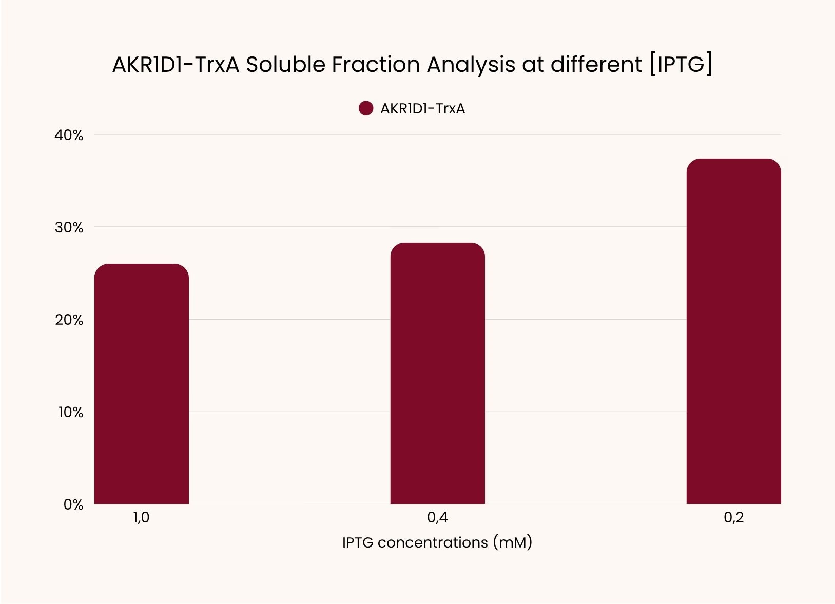
However, inconsistencies in protein quantification and sample normalization limited the accuracy of these estimates.
Check our results here.
| Week | Relevant Experimental Work |
|---|---|
| 29 | Initial expression of ci2350–His and ci2350–MBP, no visible overexpression of ci2350-MBP |
| 30 | Testing new expression conditions for ci2350-His and ci2350-MBP, confirmation of overexpression via SDS-PAGE |
| 31 | Expression comparison at different temperatures and IPTG concentrations. Collection and storage of ci2350-MBP for planned enzymatic activity assessment |
| 36 | Comparison of AKR1D1-His, SUMO and TrxA soluble fractions |
| 38 | Optimization of AKR1D1-TrxA expression by lowering IPTG concentrations |
Learn
After having identified our problem and chosen the strategy to continue, we managed to successfully clone our construct into E. coli, and non-mutated sequences were carried forward for protein expression.
After reflecting on the experiments we realised that the initial problem was insufficient digestion time, our hypothesis is strengthen by Week 30 experiments: AKR1D1-His was re-incubated with digestion enzymes and successfully cloned into E. coli.
Developing a Reliable Enzyme Activity Assay
Challenge
After producing our candidate enzymes, we needed to determine whether they could support the intended testosterone-transforming reaction. Our initial assay measured the consumption of the cofactorA helper molecule an enzyme needs to do its job. NAD(P)HA molecule that carries electrons in cells. The enzymes use it as a cofactor, and it absorbs light at 340 nm while its oxidized form does not. spectrophotometricallyUsing a spectrophotometer, which measures how much light a sample absorbs.. The development of this assay was complicated by substrate precipitation, inconsistent absorbanceHow much light a sample absorbs at a given wavelength. measurements, inadequate signal detection, and background cofactor consumption. These limitations required several rounds of troubleshooting before the measurements could be interpreted reliably.
Design
Building from existing literature (Arp et al., 2025; Penning, 2023), we developed a continuous spectrophotometric assay based on monitoring the decrease in NAD(P)H absorbance at 340 nm.
We designed assays with different enzyme concentrations to investigate whether the absorbance signal depended on the amount of enzyme-containing preparation. The following iterations focused on improving substrate preparation, selecting appropriate measurement conditions, and introducing controls to distinguish enzyme-associated activity from background reactions.
Build
We investigated activity for the following constructs:
- ci2350–MBP
- AKR1D1–SUMO
- AKR1D1-TrxA
- AKR1D1-His
Expand this section and find out about the iterative cycles!
Test
Initial enzyme-containing samples showed decreases in NAD(P)H absorbance. However, similar decreases were also observed in empty-vector preparations, preventing enzyme-specific interpretation of the signal.
Bradford analysisA color-based test for measuring how much protein is in a sample. subsequently revealed substantial protein concentrations in the empty-vector controls, including 0.926 mg/mL in the induced control, compared with 0.695 mg/mL in the ci2350–MBP double-mutant preparation.
These findings suggest non-specific protein recovery or possible carryover during purification, although the cause was not established. When the assays were repeated using ci2350–MBP and its double mutant, no clear NADPH consumption signal was observed. Prolonged storage of the enzyme preparations at 4 °C was considered a possible explanation.
Check our results here.
| Week | Relevant Experimental Work |
|---|---|
| 32 | Exploratory testosterone reduction assays with ci2350-MBP and AKR1D1-His, inconclusive results due to technical and sample handling errors. |
| 35 | Testosterone reduction assay troubleshooting using AKR1D1-SUMO, AKR1D1-His and AKR1D1-TrxA. NADPH calibration curve was introduced, assay results remained inconclusive. |
| 36 | Preparation and purification of ci2350–MBP, mutant, and empty-vector controls |
| 37 | Bradford quantification showed high protein concentration in empty-vector controls. Assay results were inconclusive as control samples showed use of NADPH as a cofactor. |
| 38 | Assays performed on ci2350-MBP and ci2350-MBP double mutant. We continued troubleshooting but NADPH measurements remained inconclusive |
| 39 | Background NADH consumption is identified as a major limitation of the assay |
| 40 | Troubleshooting testosterone-reduction assays, introduction of substrate-free negative control, NADH measurements remained inconclusive |
Learn
The experiments demonstrated that NAD(P)H consumption alone could not establish enzyme-specific testosterone conversion because background cofactor consumption, uncertain sample composition, and variability between experiments limited the interpretation of our results. The available measurements could not confirm the intended testosterone transformation or any reduction in androgenic activity. Further testing should include Mass SpectrometryMeasures the mass of molecules very precisely, so it can tell testosterone apart from its reduced product. and/or Androgen Receptor AssayCells carrying the androgen receptor and a reporter gene that switches on when a molecule activates the receptor. to confirm product formation and any changes in androgenic activity. Alongside this, our enzyme activity model of the R215A + V263I double mutant will serve as a predictive layer, helping us to understand the enzyme's preference between the two steroids (Results).
References
- Arp, G., Jiang, A. K., Dufault-Thompson, K., Levy, S., Zhong, A., Wassan, J. T., Grant, M. R., Li, Y., Hall, B., & Jiang, X. (2025). Identification of gut bacteria reductases that biotransform steroid hormones. Nature Communications, 16(1). https://doi.org/10.1038/s41467-025-61425-6
- Penning, T. M. (2023). Steroid 5β-reductase (AKR1D1): Purification and characterization. Methods in Enzymology, 689, 277–301. https://doi.org/10.1016/bs.mie.2023.04.012
- Shutevska, K., & Nestorovska, A. K. (2024). Cloning, expression, and purification of recombinant AKR1D1 for therapeutic applications. Acta Pharmaceutica. https://doi.org/10.2478/acph-2025-0003